EZC 2023: Meeting Recap
Meeting Report: 12th European Zebrafish Meeting, Kraków Poland, July 9th-13th 2023
By
Florence Marlow, Icahn School of Medicine, Mt. Sinai Hospital, New York and North American Representative, International Zebrafish Society.
and
Emily Noel, School of Biosciences, University of Sheffield and Member, European Zebrafish Society Steering Committee.
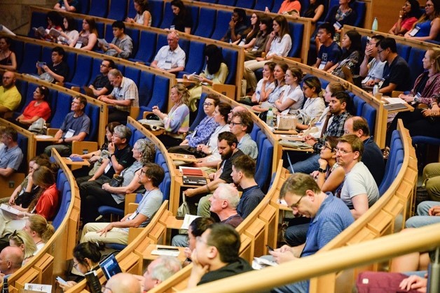
In July 2023, around 500 zebrafish researchers arrived in the beautiful Polish city of Kraków for the 12th European Zebrafish meeting. The meeting was held in the fantastic Auditorium Maximum of Jagiellonian University (see photos). The meeting was not only a forum to share, listen to, and discuss world-class science, but also a welcome opportunity to reconnect with former colleagues and friends in the shoal. For those of you who couldn’t make the meeting (and those of you who could, but would like to reminisce), with the help of the session chairs we’ve outlined some session highlights below. But first a shout-out to those excellent talks and projects that we haven’t been able to include here due to space constraints - the meeting was absolutely jam-packed with great science!
Cell biology in a fish: From single cells to building and shaping tissues
Beginning with the egg in the Germline and Early Development session, Andreas Zaucker from Karuna Sampath’s lab combined transgenesis with time-lapse analyses to track the earliest movements of the germ plasm from the vegetal pole to the cleavage furrows. Combining imaging with 3D temporal segregation he discovered stereotypic patterns of germ plasm movement and then applied this approach to ybx1 mutants and found reduced movement of germ granules animally and then to the cleavage furrows, which may account for the reduced germ line and male bias of ybx1 mutants. Luis Hernandez-Huertas (Moreno-Mateos lab) described his PhD work developing CRISPR/Cas13d RNA knockdown to study maternal mRNA activity. Excitingly, his screen of maternal kinases uncovered a cytosolic role for Branched chain ketoacid dehydrogenase kinase (Bckdk) in regulation of zygotic gene expression at the maternal to zygotic transition.
A session on Tissue formation and Patterning featured insights into our understanding of key developmental signaling pathways including Lucy Brunt’s discovery in Steffen Scholpp’s lab that in the early embryo cytoplasmic projections called cytonemes contain Wnt5b associated with its receptor Ror2. Remarkably, Lucy showed in live imaging that the Wnt5b/Ror2 complex is transported along cytonemes, which can extend over 3 cell rows, and is transferred to a receiving cell to mediate PCP-driven convergent extension. Scott Wilcockson from Caroline Hill’s lab reported on the FGF downstream signal transducer ppErk, which exhibits heterogeneous levels in cells undergoing mesendoderm patterning. He found that activated ppErk is lost during mitosis, and this erasure contributes to the heterogenous ppErk levels in cells and acts in mesendodermal patterning. Fanny Eggler (del Bene lab) generated triple mutants of the Meteorins, secreted factors expressed within the dorsal forerunner cells of the early embryo that form Kupffer’s vesicle (also called the node), an important structure in the establishment of left-right asymmetry in vertebrates. They found that the triple loss of Meteorins causes defects in dorsal forerunner cell clustering, Kupffer vesicle formation and left-right patterning defects, revealing new factors in KV formation and LR asymmetry formation. Soraya Villaseca (Scarpa lab) showed that as trunk neural crest cells migrate, they squeeze through small spaces that lead to significant nuclear deformation, which can lead to nuclear membrane disruption and nucleoplasm leakage, as shown by a nuclear GFP transgene marker and using AI to assess the circularity of the nucleus in this analysis. Hannah Greenfeld from Daniel Wagner’s lab examined the temporal role of BMP signaling in specifying three dorsal neuronal cell types of the spinal cord. She determined that only one subtype is specified during neural tube stages, whereas the other two are specified during gastrulation, possibly when neural tissue is specified.
Two Morphogenesis and Organogenesis sessions covered a diversity of organs and featured exciting research applying quantitative tools and modelling to decipher the mechanisms of tissue morphogenesis. Iris Unterweger from Elke Ober’s lab presented exciting work that involved mapping liver cell fate using the multicolor FRAEPPLI approach. In her presentation, Iris showed that liver progenitors (hepatoblasts) can either be bipotent or unipotent giving rise to hepatocytes and cholangiocytes. The team also revealed new insights regarding the morphogenesis of the multi-lobed adult liver, identifying the origin of ventral lobe formation during postembryonic development. Additional highlights from the session included Nikolay Nikov’s work on pancreatic beta-cells. Using neural circuit tools (Campari, optogenetics, calcium imaging) they showed that a single leader cell initiates a Ca2+ wave that is then propagated to other cells. Rolling with the theme of waves, Alessandro de Simone presented beautiful data on Erk activity waves and their contribution to scale regeneration. Finally, Tony Tsai shared the cadherin code underlying spinal cord morphogenesis, an extension of the groups earlier work.
Zebrafish in Health and Disease
The advantages of using zebrafish as a model to investigate disease aetiology, pathophysiology, and as a preclinical model for drug discovery was exemplified by the variety of disease models presented at the meeting, including developmental and physiological disorders, aging and age-related diseases, neurological and psychiatric disorders and infectious disease.
In diseases of bone homeostasis two talks highlighted zebrafish models of progressive bone overgrowth disorders. Yentl Huybrechts (van Hul lab) presented the first zebrafish model of Paget’s disease of bone (PDB). PDB is a disorder characterized by the presence of hyperactive osteoclasts and focal increases of bone turnover. They targeted the sqstm1 gene, the major disease-causing gene in PDB, mutating a domain in which heterozygous variants are associated with disease in humans. Micro-CT analyses revealed defects in the vertebral column at 12 month of heterozygous zebrafish mutants, and accelerated bone turnover. Nicola Blum (Harris lab) is investigating Proteus syndrome, a rare disorder also characterized by progressive focal bone overgrowth. Proteus syndrome patients have mosaic somatic activating mutation in AKT1. Mosaic expression of the corresponding zebrafish Akt1 mutation in F0 embryos results in localised embryonic bone overgrowth, and injury-dependent progressive bone overgrowth phenotypes in adults.
A variety of sensory and neurological disorders were presented, highlighting the breadth of current work in zebrafish models of neurological disease. Nerea Montedeoca from Berta Alsina’s lab presented data investigating sall1a, the ortholog of human SALL1 which is mutated in Townes-Brocks syndrome (TBS). TBS is an autosomal dominant multisystemic disorder including sensorineural hearing loss, and Nerea showed that the sall1a heterozygotes and homozygous mutant larvae have reduced numbers of functional mature hair cells and balance defects. Yuko Nishiwaki (Masai lab) presented PDE6c mutants as a zebrafish model of photoreceptor degeneration. PDE6c is a cone cell-specific subunit of PDE6, and PDE6c mutants exhibit cone cell degeneration in larvae . Yuko investigated the role of calcium signalling in cone cell degeneration in PDE6c mutants and found elevated levels of calcium in photoreceptors, which could be rescued by cone-specific knockdown of CNG channels. Paul Kasher presented a zebrafish model of Aicardi-Goutières syndrome (AGS), an inherited inflammatory brain disease which can be caused by loss of the viral restriction factor SAMHD1. Loss of samhd1 in zebrafish larvae induced substantial, but variable, upregulation of interferon stimulated genes expression and exhibit microcephaly, cell death in the brain, and an increased susceptibility to brain haemorrhage upon cholesterol dysregulation. They also describe significant dysregulation of cholesterol biosynthesis in both AGS patients and samhd1 zebrafish mutant larvae, although surprisingly the direction of dysregulation is opposing. Petronella Kettunen described zebrafish knockout models of the quaking (QKI) gene, an RNA metabolism gene involved in myelination and which is downregulated in the brains of schizophrenic patients. Zebrafish qkib (the ortholog of the QKI variant downregulated in schizophrenia patients) mutants exhibit hallmarks of increased anxiety, and altered cortisol secretion, a physiological response to chronic stress. The qkib mutants also exhibit structural and biochemical differences, that correspond with defects observed in schizophrenic patients.
Defects in vascular integrity can occur in a variety of diseases. Victor Tapia (Kasher lab) presented his work on the role of cholesterol dysregulation in intracerebral haemorrhage (ICH) in a statin model, where decreased cholesterol synthesis leads to increased ICH risk. They propose a novel pathway where antimicrobial interferon signalling and cholesterol dysregulation lead to ICH. Emilia Seta (Prajsnar lab) investigated the mechanism of Ponatinib, a chemotherapeutic agent known to induce adverse vascular toxicity. Through analysis of tail and cerebral vasculature in Ponatinib-treated embryos, Emilia demonstrated that Ponatinib induces increased vascular permeability and vasoconstriction. Maximilian Breuer (Abdelilah-Seyfried lab) showcased his work using zebrafish models to understand endothelial ageing. Hutchinson-Gilford Progeria Syndrome (HGPS) is a rapid ageing disorder caused by mutations in LaminA which led to nuclear accumulation of Progerin. Max generated a Progerin overexpression transgenic zebrafish as a model of rapid ageing, and examined endothelial phenotypes, reporting that ageing altered the structure of endothelial cell nuclei, morphology, and their ability to respond to mechanical stimuli.
The strengths of zebrafish to model infection was highlighted by the variety of infection models on display. Serge Mostowy presented a new zebrafish model to study Shigella infection, and the role of Septins in infection control. His work revealed new insights into Shigella-phagocyte interactions, and has exploited this new model to study trained immunity in vivo. Beatriz Novoa gave an overview of viral infection research in zebrafish, including recent studies using Sars-Cov2 proteins as immune triggers, and the use of zebrafish to screen for compounds with anti-inflammatory activity against Sars-Cov2 trigger. Magdalena Widziolek (Chadzinska lab) described her investigation in Porphyromonas gingivalis (Pg) which causes periodontitis in humans and has been linked with comorbidities. A zebrafish Pg infection model reveals Pg induces vascular permeability through degradation of endothelial adhesion proteins, and neuroinflammation, providing a new model to further explore brain pathologies that have linked with this gum disease pathogen.
Technological Advances
Exciting developments in imaging approaches and analyses, novel reporter and transgenic lines, modified genetic approaches, and detailed genomic resources were on show not only in the Emerging Technologies session, but across the meeting. Advances in imaging and image analysis included data from Katerina Makarova (Magorżata lab), showcasing how X band Electron Paramagnetic Resonance can be applied in live zebrafish larvae to detect, identify and quantify free radicals. Katarina demonstrated EPR detection of melanin radicals and the use of spin probe EPR to detect changes in membrane fluidity and permeability, demonstrating the impacts of hydrogen peroxide or DMSO on membrane fluidity in larvae. This approach has thus far been tested on embryos from 3dpf to 5dpf. Julia Noeth (Scholz lab) has developed a novel system for automated label-free imaging to detect vascular disruption in larvae, taking advantage of the ability to visualise blood cell movement through the vasculature. In this approach, short recordings of blood flow through the vasculature are created, and subsequently anything in the image that is not moving is removed, resulting in a subtraction image where only the vasculature is retained. Comparative analysis of subtraction images with vascular transgenic lines can categorise spatiotemporal patterns of blood flow in developing vessels, providing greater details about the relationship between vasculogenesis, lumenisation and flow. Graham Lieschke presented work from his group using cutting-edge lattice light sheet imaging to resolve the nuclear envelope in migrating neutrophils, and bespoke algorithms for segmenting nuclei for morphometric analysis of nuclear dynamics in migrating neutrophils. Their data reveals high levels of plasticity in nuclear shape, with links between nuclear shape and neutrophil velocity, and detailed analysis of nuclear position challenges current models of how the nucleus positions and influences inflammation-induced cell migration.
On show was the continued development of novel reporter and transgenic lines. Helen Eachus (Soojin lab) presented their new transgenic zebrafish line star:bPAC which allows elevation of glucocorticoid levels through optogenetic activation of adrenal cells. Costantino Parisi (Winata lab) has identified a region on chromosome 7 that acts as an enhancer specifically in trabecular cardiomyocytes. Using this enhancer they generated a reporter line which demarks trabecular cardiomyocytes in the ventricular wall even prior to the onset of trabeculation. Staying in the cardiovascular system, Christian Helker presented new Apelin reporter transgenics to provide more detailed analyses of apelin expression dynamics in the developing vasculature, using either Venus-PEST (destablilised GFP with a short half-life), or superfolding GFP. Christian also presented a new zic2b:mCherry roof plate reporter. Konstantinos Kalyviotis presented GenEPi, a genetically-encoded fluorescent reporter for non-invasive optical monitoring of Piezo-1 dependent cellular mechanotransduction. Martin Distel presented new tools to distinguish neutrophil maturation grades, including Tg(lysC:CFPNTR) and Tg(BACmmp:CitrineCAAX) lines to distinguish immature and mature neutrophils respectively. Finally, Iris Unterweger (Ober lab) described their multispectral cell labelling tool FRAEPPLI, allowing them to map cell hepatic cell fates. FRAEPPLI features both nuclear and membrane-localized fluorophores, is developed in the GAL-UAS system, and is compatible with SeeDB2 clearing.
This year featured several developments in conditional gene regulation, Cre-mediated recombination, and genome engineering. Maura McGrail has developed Cre/lox tools for conditional epigenetic regulation. Her team has applied GeneWeld targeted knockin technology to generate a floxed conditional allele of the histone deacetylase Hdac1. Komali Valishetti reported a single vector system for cell-type specific Cre recombinase cell lineage tracing that allows either chemical or light control of recombinase efficiency. This approach can facilitate introduction of sparse labelling in specific cell populations or tissues. Ramy Elsaid (Hernandez lab) presented their transgenic light-inducible Magnets-based Cre/lox system, emplying a split Cre that functions across tissues and developmental stages for non-invasive blue light-controlled tracking of cell fate and migration in vivo. Luis Hernandez-Huertas (Moreno-Mateos lab) decribed their use of an optimised CRISPR/Cas13d system for RNA targeting to study maternal mRNA function.
Community resources and initiatives
The expanding use of omics technologies at scale led several researchers to present new genomic resources for the community. Emre Yaksi presented his team’s Atlas of Zebrafish Transcriptome Encephalic Cytoarchitecture (AZTEC). AZTEC was developed through spatial trancriptomics of the zebrafish telencephalon at sub-cellular resolution. Notably, using this approach they also identified that a significant number of transcripts were found in the extracellular space. Through analysis of 99 genes from AZTEC to stratify cell types, along with integration of AZTEC with published scRNA-seq datasets, they revealed previously uncharacterized forebrain nuclei and identified cortical regions conserved across vertebrates. Merlin Lange (Loic group) has developed Zebrahub, an online resource providing single-cell sequencing datasets that encompass 10 stages of embryonic development, from 10 hours to 10 days post fertilisation. The Zebrahub atlas also incorporates a computational pipeline for tracking cell fate via light sheet microscopy. The Zebrahub team demonstrated the power of this multi-modal approach by fate mapping Neuro-Mesodermal Progenitors during embryogenesis. Finally, Shawn Burgess’s team is working towards an updated telomere-to-telomere zebrafish genome assembly.
Building upon broader community awareness and commitment to environmental sustainability the Krakow meeting featured a plenary session devoted to environmental impacts of research, using zebrafish to understand physiological changes and reponses to environmental stressors. This included the notable talk by Florence Kermen on neural function during heat stress – something we could all relate to with the recent heat waves - as well as community initiatives such as the first Zebrafish Lab Waste Audit spearheaded by Maximillian Breuer and Viviana Vedder and Malgorzata Grodzinska-Jurczak’s work to integrate citizen science and science communications to address single-use-plastic pollution.
Young Researchers Committee (YoRC) session
This iteration of the European Zebrafish Meeting hosted the first in-person YoRC session. Konstantinos Kalyviotis, the YoRC chair, gave a great overview of the aims and objectives of the committee. The YoRC was established in 2021, with the goal of connecting early career researchers, to foster information and knowledge exchange, and to showcase the research carried out by early-career scientists. The session kicked off with two longer presentations. Ximena Soto (Papalopulu lab) presented live imaging work looking at gene expression oscillation in cell-fate decisions of neural progenitors, through generation of a novel Her6:Venus transgenic line. She uncovered temporal variations in oscillation noise and dynamics across neuronal development, and identified miR-9 as a dampener of oscillatory noise. Diana Kaszás (Enyedi lab) showcased her work on purinergic and calcium signalling pathways in tissue damage. She used a variety of transgenic biosensors, including the GrabATP to visualise extracellular ATP release, and gCamp7s-NLS to assess calcium signalling, in a larval tail-injury model. Diana found that extracellular ATP induced calcium increase during injury, potentially through the P2X7 receptor, to promote subsequent wound closure.
A series of excellent flash talks were presented by ECRs, including MSc students, and the session rounded off with a presentation from Anja Glenk on regaining confidence and joy in research. Anja highlighted important issues surrounding mental health in academia, including the value of discussing mental health stressors and impacts together as a community. She also suggested positive actions we can take to support our own mental wellbeing, including tips on positive rephrasing of negative thoughts, and building and supporting self-confidence. We recommend checking out her website for more information https://anja-glenk.com/.
Recognizing achievements, leaders, and role models in our community
Our vibrant community was built upon and continues to thrive due to the combination of stellar science, generosity, and collegiality of our members, both long time members and more recent converts. In recognition of these attributes three awards were given at the EZM meeting in Krakow. The Christiane Nüsslein-Volhard Award, named in honor of the the outstanding contributions of Nobel prize winner Christiane Nüsslein-Volhard to the field of genetics, is awarded by the European Zebrafish Society in recognition of outstanding achievements in Zebrafish research. Outstanding achievements include substantial advances in technology within the Zebrafish field, scientific advances published in excellent papers, or outstanding contribution to the infrastructure and funding resources zebrafish community. This year’s Christiane Nüsslein-Volhard Award recipient was Dr. Mary C. Mullins of the University of Pennsylvania USA, in recognition of Mary’s outstanding scientific contributions and unwavering commitment to advancing our understanding of genetics and for her ground-breaking work on dorsal ventral patterning of the embryo and maternal control of development using zebrafish. Mary is a leader in our field both scientifically and as a mentor through her direct contributions to training scientists in her own lab and as Co-Director and faculty in the Zebrafish Course held annually in Woods hole, and for her community leadership as the first vice president of the International Zebrafish Society and second president, and for her service on study sections as a reviewer and chair, and as editor of PloS Genetics, positions that Mary leveraged to influence the broader scientific community and advocate for greater acceptance and recognition of the zebrafish as a major vertebrate model for genetics research. Mary initially trained as a biochemist before performing pioneering work to establish the mutagenesis methods that made the large-scale genetic screens possible and led to the identification of more than a thousand zebrafish mutants disrupting early development. In her lab at UPenn, Mary combined her biochemistry background and zebrafish genetics to dissect the contributions of BMP signaling to patterning the early embryo provided evidence that heterodimers of ligands and receptors are required for signaling among other important insights into BMP contributions to development. In parallel, Mary led the first large-scale maternal-effect screens in a vertebrate, providing access to the genetic factors regulating early oocyte development and the earliest events of embryogenesis. In her talk, Mary expressed gratitude to the field and community for being collaborative and collegial and she thanked and emphasized the fun and rewarding experience she has had working with talented postdoctoral fellows and students. Mary emphasized the challenges and rewards of the maternal-effect screens and highlighted the exciting mutants and genes that emerged as a result, sharing vignettes of each student and fellows’ contributions. Mary also shared her fascination with morphogen gradients, including how they are shaped and formed and how they contribute to development and then highlighted the key discoveries made by her group, including the importance of heterodimers, as well as new discoveries made using a combination of genetic and computational approaches to test and ultimately revise the models for BMP gradient formation during dorsal ventral patterning.
The George Streisinger Award, named in honor of the founding father of zebrafish research, George Streisinger (1927-1984). George Streisinger established a zebrafish research colony at the University of Oregon in the 1970s and early 1980s and developed the first methods for mutagenesis and mutant screening with the goal of studying the development of the nervous system through genetic analysis. George Streisinger’s enthusiasm for the zebrafish and its potential for understanding vertebrate development inspired numerous researchers including his colleagues at the University of Oregon and around the world, who after Dr. Streisinger’s untimely death in 1984, stewarded development of the still-nascent model organism to establish the foundation of today’s thriving community. The International Zebrafish Society (IZFS) established the George Streisinger Award to honor Dr. Streisinger's outstanding contribution to the field and to recognize a senior investigator who’s sustained, foundational, and transformative work has benefited and strengthened the zebrafish field. This year’s recipient of the George Streisinger Award was Dr. John Postlethwait of the University of Oregon, USA, in recognition of his sustained and numerous transformative contributions to the field, including being one of the early stewards of the field, for his monumental contributions to understanding the complex zebrafish genome, including the radiation hybrid and linkage maps that made pinpointing the chromosomal position of genetic lesions possible, as well as for his ground-breaking work on the mechanisms of sex determination. John’s talk was a masterclass in giving an award talk. John trained at Case Western Reserve University prior to joining the faculty at the University of Oregon, where he knew and worked closely with George Streisinger. In his three-part talk, John highlighted discoveries of Streisinger that made his own work and the work of others possible. He talked about George and Charline Walker’s haploids, half-tetrads, and George’s work on phage and the genetic code which made use of CRISPR possible and allowed for the important work of the Undiagnosed Disease Network (UDN) to provide genetic insight into the causes of rare diseases. He highlighted how George’s double haploids were critical to generating the genetic maps for AB and Tubingen strains used for the 2013 Zebrafish genome. Finally, he talked about George and sex chromosomes, that George had noticed the sex ratio distortions that inspired John's lab to map sex like mapping a mutant, which ultimately led him to the wild where he recovered the Nadia and Cooch Behar strains that revealed loss of the ZW sex determination system in the inbred lab strains. His talk highlighted his foundational studies as well as exciting new work toward identifying the elusive sex determinant, and expressed his gratitude to George, Charline, the people he has worked with and the field.
The Chi-Bin Chien Award was established by the zebrafish research community and the International Zebrafish Society (IZFS) in memory of Dr. Chi-Bin Chien. Chi-Bin Chien (1965-2011) was Professor of Neurobiology and Anatomy at the University of Utah and served the international zebrafish community in numerous ways, including his service as Director of the Zebrafish Neural Development and Genetics Course at the Woods Hole Marine Biological Laboratory and as an organizer of the International Conference on Zebrafish Genetics and Development. Chi-Bin Chien was beloved for his enthusiasm and love for scientific discussions and the benefits of open communication among scientists, his devotion to mentoring and supporting the development of young scientists, and for his collaborative and generous spirit. The award named in Chi-Bin’s honor recognizes outstanding graduate students, postdoctoral trainees, or recently appointed faculty members from any country who have made significant contributions to the zebrafish field and who embody Chi-Bin’s generous and open nature. Dr. Peter Fabian, a former fellow in Gage Crump’s lab and now Assistant Professor at Masaryk University in Czechia received the Chi-Bin Chien Award at the meeting in Krakow for his groundbreaking zebrafish research and efforts tto build connections between the zebrafish community in Czechia, other scientists, and with other countries in Europe. Peter’s talk featured his “pirate work” on endoderm while using scRNAseq and lineage tracing tools to study neural crest during his postdoctoral days, which led to the unanticipated discovery that the anterior most endoderm contributes to the pituitary, which was previously thought to develop from the ectodermal placode. Based on evolutionary comparisons, including with amphioxus, Peter thinks this endodermal contribution is an ancient vestigial feature. In the second half of his talk Peter focused on neural crest and dermal fibroblasts. He shared exciting recent work showing that a gene associated with black bone disease, Alkaptonuria, which in humans leads to discoloration of body fluids and collagen rich tissues, including blackening of the bones. Significantly, mouse models of Alkaptonuria fail to recapitulate the blackening of the bones, hindering studies of the disease and identification of therapeutics. Excitingly, upon returning from a Covid-19 induced hiatus Peter returned to the lab to discover that his zebrafish mutants fully recapitulate the human disease and thus position Peter and his new lab to provide needed mechanistic insight and potentially uncover therapeutic strategies for this disease. By the way, if you are in the market for an exciting training opportunity with an engaging scientist who we can confirm can also tear up the dance floor, Peter is hiring.
The Keynote lectures are a highlight of every zebrafish gathering and the 12th EZM keynote delivered by Dr. Magdalena Zernicka-Goetz, Professor of Development and Stem Cells at the University of Cambridge and Bren Professor of Biology and Biological Engineering at the California Institute of Technology, was no exception. Dr. Zernicka-Goetz highlighted her lab’s beautiful and transformative work on patterning the early mouse embryo and using the rules learned from the embryo to coax stem cells, from mouse and human, to organize into assemblies that resemble the early embryo, thus opening the door for functional studies of the earliest events in development of the human embryo. Dr. Zernicka-Goetz shared spectacular images as she walked us through her scientific journey from embryology studies to engineering embryo-like assemblies from pluripotent stem cells, pointing out influences and inspiration from zebrafish work and the parallel and collaborative work of others.
Gratitude and next gathering of the shoal
On the final full day of the meeting, the local organisers had arranged for a conference banquet to be held in the delightful Folwark Zalesie, a 19th century Manor house in the countryside outside Kraków. After being welcomed with drinks and enjoying dinner, delegates were treated to (and encouraged to join in with!) a display of traditional Polish dancing running on into the warm Kraków evening. We’ve included a few photos of the meeting below, but the full gallery can be found here.
With the 12th European Zebrafish meeting behind us, we would like to extend a huge thank you to the International Advisory Board (Przemko Tylzanowski, Stefan Schulte-Merker, Lilianna Solnica-Krezel, Antonella Forlino, Koichi Kawakami, Jacek Kuznicki, Graham Lieschke,Tomasz Prajsnar, Mate Varga, and Cecilia Lanny Winata), the Krakow EZM 2023 local meeting organizers (Przemko Tylzanowski, Piotr Podlasz, Marta Migocka-Patrzalek , Magdalena Chadzinska, Tomasz Prajsnar, Krzysztof, Anna Sarosiak), the session chairs (an extra thanks for their assistance in constructing this report), our local hosts at Jagiellonian University for the stellar conference support and for feeding us so well, the conference sponsors, and all who participated, shared their work, exchanged ideas, and brought your enthusiasm to the sessions and the dance floor.
The next opportunity to flip your fins and shoal with our international community will be the 18th International Zebrafish Conference in the stunningly beautiful and tranquil Kyoto, Japan from August 17-21, 2024. Thank you in advance and good luck to the 2024 conference organizing committee (Corinne Houart, Hitoshi Okamoto, Miguel Luis Concha, Hiromi Hirata, Tohru Ishitani, Florence Marlow, Didier Stainier, Han Wang, and Karina Yaniv). Mark your calendars and look out for additional details about the venue and key dates at izfs.org. We look forward to swimming with you all in Kyoto!
|
Delegates at the main auditorium at the EZM |
Traditional dancing at the conference banquet |
|
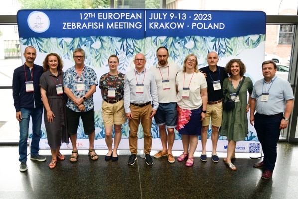
The EZM Organizing Team |
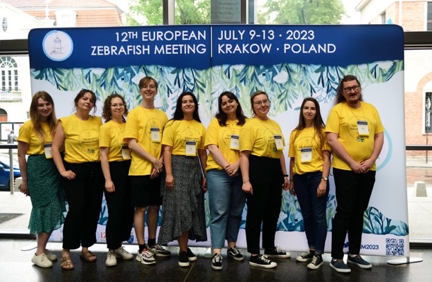
EZM Volunteer Crew |
|
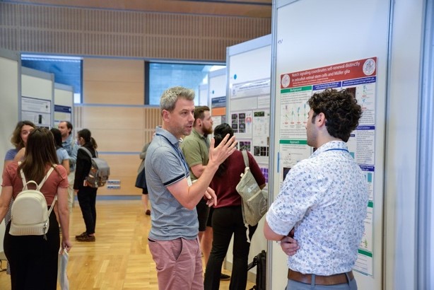
Poster sessions at the EZM |
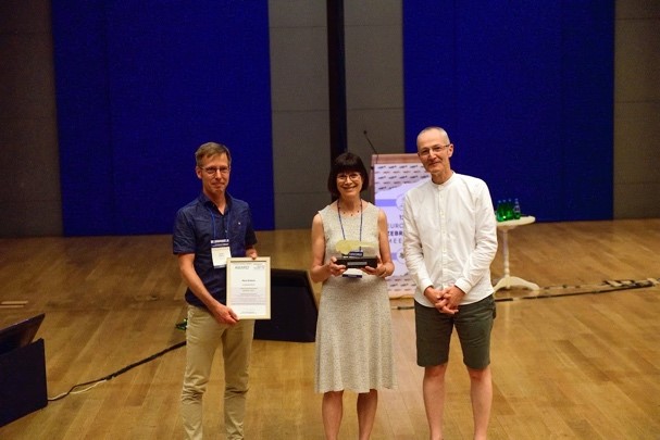
Mary Mullins receiving the CNV Award |
|
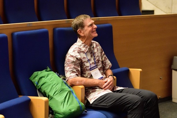
John Postlethwait, winner of the George Sreisinger Award |
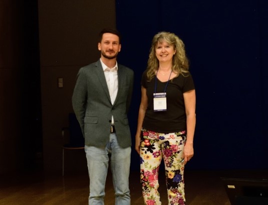
Peter Fabian, winner of the Chi-Bin Chien Award with IZFS President Corrinne Houart |